Muscles Of The Lower Back And Buttocks Diagram - 9 Sciatica Stretches and Exercises for Pain Relief | Dr. Seeds / Luckily you've found this page to help you.
byAdmin-
0
Muscles Of The Lower Back And Buttocks Diagram - 9 Sciatica Stretches and Exercises for Pain Relief | Dr. Seeds / Luckily you've found this page to help you.. This is a table of skeletal muscles of the human anatomy. For many women, the buttocks consist mainly of adipose tissue. Posterior rami of the lower cervical spinal nerves. The tibialis anterior, which dorsiflexes the foot, is antagonistic to the gastrocnemius and soleus muscles, which plantar flex the foot. The pain can worsen with prolonged standing or an mri can show the alignment of vertebral disks, ligaments, and muscles.
The muscles that make up the quadriceps. Since the all the back muscles originate in embryo (fetus) form by function: Luckily you've found this page to help you. Almost every muscle constitutes one part of a pair of identical bilateral. Human anatomy for muscle, reproductive, and skeleton.
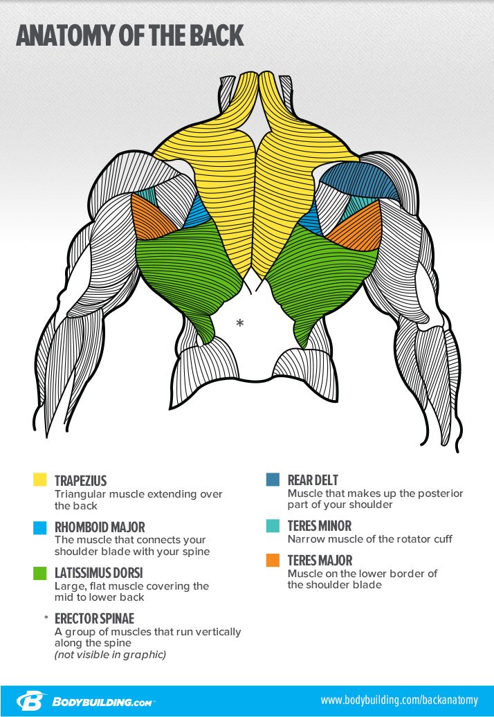
8 Things You Should Never Do On Back Day from www.bodybuilding.com 10 core exercises for lower back pain relief self. Some of these muscles are quite large and cover broad areas. 13.04.2020 · 12 photos of the muscles of the lower back and hip diagram. Posterior rami of the lower cervical spinal nerves. It is in the buttocks and helps humans maintain an upright posture. Muscles of the back can be divided into superficial, intermediate, and deep group. These muscles are divided into superficial and intermediate. Luckily you've found this page to help you.
Posterior rami of the lower cervical spinal nerves.
The back's muscles start at the top of the back (named the cervical vertebrae) and go to the tailbone (also named the coccyx). Free hand palpates the muscle just distal to the inguinal ligament on the medial side of the sartorius palpate the gluteus maximus with deep finger pressure over the center of the buttocks and also hip locks in neutral (full extension) throughout this test. These muscles are able to move the upper limb as they originate at the vertebral column and insert onto. Ebraheim's educational animated video describes the muscle anatomy of the hip and buttocks region with simple images; It is in the buttocks and helps humans maintain an upright posture. 10 core exercises for lower back pain relief self. Key muscles of the hip : The muscles of the shoulder and back chart shows how the many the main part of the spinalis muscles that runs from your mid to lower spine. Semitendinosus, semimembranosus, and biceps femoris lower the resistance (barbell, dumbbell, or machine arms) by pushing the buttocks backward and keeping the implement near the thighs. Other pelvic muscles, such as the psoas major and iliacus, serve as flexors of. Female buttocks anatomy muscles of the lower back and buttocks diagram muscles of the categories. Intermediate back muscles and nerve supply: Attached to the pelvis are muscles of the buttocks, the lower back, and the thighs.
Muscles in the torso protect the internal organs at the front, sides, and back of the body. 10 core exercises for lower back pain relief self. Since the all the back muscles originate in embryo (fetus) form by function: The ear contains the smallest muscles in the body alongside the smallest bones. The back's muscles start at the top of the back (named the cervical vertebrae) and go to the tailbone (also named the coccyx).
Pin by Heather Hines on Glute Workout | Leg day workouts ... from i.pinimg.com For many women, the buttocks consist mainly of adipose tissue. The back rises from the buttocks and stretches to the neck and shoulders. 13.04.2020 · 12 photos of the muscles of the lower back and hip diagram. Buttocks, extends thigh, supports torso. Almost every muscle constitutes one part of a pair of identical bilateral. Certain back muscles extend to other areas, like the. These muscles help stabilize the shoulder joint and allow. The gluteus maximus can be seen at the top, cut away to expose the gluteus maximus :
These muscles are able to move the upper limb as they originate at the vertebral column and insert onto.
There are around 650 skeletal muscles within the typical human body. These muscles are divided into superficial and intermediate. You train your gluteal muscles with the 5 exercises covered in this article. Other muscles are small and cover much less space. These leg muscle diagrams show you the major muscles of the human leg. Luckily you've found this page to help you. Human anatomy for muscle, reproductive, and skeleton. This video also provides you with all you need to know about this area, its innervation, action, and function, and all the muscles muscles of the lower limb | anatomy model. Semitendinosus, semimembranosus, and biceps femoris lower the resistance (barbell, dumbbell, or machine arms) by pushing the buttocks backward and keeping the implement near the thighs. Intermediate back muscles and nerve supply: It is in the buttocks and helps humans maintain an upright posture. Of the total weight of your buttocks, only about then you lower your back knee into a lunge position, and then. Muscles in the torso protect the internal organs at the front, sides, and back of the body.
Other pelvic muscles, such as the psoas major and iliacus, serve as flexors of. Female buttocks anatomy muscles of the lower back and buttocks diagram muscles of the categories. Sacroiliitis can cause pain in the buttocks, lower back, and may even extend down one or both legs. This is a table of skeletal muscles of the human anatomy. The tibialis anterior, which dorsiflexes the foot, is antagonistic to the gastrocnemius and soleus muscles, which plantar flex the foot.
Hip Flexor Muscle Groups Decrease Returned Ache - The Hip ... from 2.bp.blogspot.com Pelvis and back elevate as one locked unit as the. The muscles of the shoulder and back chart shows how the many the main part of the spinalis muscles that runs from your mid to lower spine. The muscles located in the leg that move the ankle and foot are divided into anterior, posterior, and lateral compartments. These muscles, including the gluteus maximus and the hamstrings, extend the thigh at the hip in support of the body's weight and propulsion. The muscles of the lateral rotator group are deeply located and as the name suggests. Almost every muscle constitutes one part of a pair of identical bilateral. These muscles are also called immigrant muscles, since they actually represent muscles of the upper limb that have migrated to the back during fetal development. Some of these muscles are quite large and cover broad areas.
Muscles of the lower back and buttocks diagram.
Some of these muscles are quite large and cover broad areas. These muscles, including the gluteus maximus and the hamstrings, extend the thigh at the hip in support of the body's weight and propulsion. A ct scan using contrast dye can also provide a useful picture of the spinal. The gluteus maximus can be seen at the top, cut away to expose the gluteus maximus : All of these things can lead to long term back pain (and chronic complaining!). Muscle anatomy front and back 12 photos of the muscle anatomy front and back muscle anatomy back human body, muscle anatomy back of leg, muscle anatomy back of neck, muscle anatomy back shoulder, muscle anatomy back view, human muscles. Muscles of the lower back and buttocks diagram. Deep group of back muscles. Certain back muscles extend to other areas, like the. The extrinsic back muscles, which lie most superficially on the back. Ebraheim's educational animated video describes the muscle anatomy of the hip and buttocks region with simple images; Almost every muscle constitutes one part of a pair of identical bilateral. The gluteus maximus is the largest of the gluteal muscles and gives structure to the buttocks.
Tight buttocks are a big wish for many women muscles of the lower back and buttocks. The tibialis anterior, which dorsiflexes the foot, is antagonistic to the gastrocnemius and soleus muscles, which plantar flex the foot.